You are looking for information, articles, knowledge about the topic nail salons open on sunday near me 단백질 전기 영동 결과 해석 on Google, you do not find the information you need! Here are the best content compiled and compiled by the https://chewathai27.com/to team, along with other related topics such as: 단백질 전기 영동 결과 해석 전기영동 결과 보는법, 전기영동법, dna 전기영동, Protein electrophoresis, 2차원 전기영동, Immunofixation electrophoresis, 전기영동 종류
단백질 전기 영동 결과 해석
- Article author: www.sangji.ac.kr
- Reviews from users: 29815
Ratings
- Top rated: 4.8
- Lowest rated: 1
- Summary of article content: Articles about 단백질 전기 영동 결과 해석 전기영동:용액에 전류를 통하면 용액중의 하전 입자는 양극 또는 음극으로 이동 … 1)단백 전기영동 (Protein EP) : 검체 혈청, 뇨, CSF … 단백전기영동 결과 해석. …
- Most searched keywords: Whether you are looking for 단백질 전기 영동 결과 해석 전기영동:용액에 전류를 통하면 용액중의 하전 입자는 양극 또는 음극으로 이동 … 1)단백 전기영동 (Protein EP) : 검체 혈청, 뇨, CSF … 단백전기영동 결과 해석.
- Table of Contents:

단백질 sds-page 결과값 보는 법 좀(사진有) > BRIC
- Article author: www.ibric.org
- Reviews from users: 41597
Ratings
- Top rated: 4.4
- Lowest rated: 1
- Summary of article content: Articles about 단백질 sds-page 결과값 보는 법 좀(사진有) > BRIC 제가 단백질 전기영동 실험한 결과인데요어떤 분자량의 단백질들이 있는 건지요??실험 결과 보는 법을 잘 모르겠네요..맨 왼쪽이… …
- Most searched keywords: Whether you are looking for 단백질 sds-page 결과값 보는 법 좀(사진有) > BRIC 제가 단백질 전기영동 실험한 결과인데요어떤 분자량의 단백질들이 있는 건지요??실험 결과 보는 법을 잘 모르겠네요..맨 왼쪽이… 제가 단백질 전기영동 실험한 결과인데요어떤 분자량의 단백질들이 있는 건지요??실험 결과 보는 법을 잘 모르겠네요..맨 왼쪽이…BRIC, 생물학연구정보센터, 단백질, 전기영동, 결과도출
- Table of Contents:
 Read More
단백전기영동
- Article author: labtestsonline.kr
- Reviews from users: 23811
Ratings
- Top rated: 3.7
- Lowest rated: 1
- Summary of article content: Articles about 단백전기영동 단백전기영동 검사는 어떻게 활용되며, 검사는 언제 시행하는지, 검사 결과의 의미는 무엇 … 단백전기영동(Protein Electrophoresis); 면역고정전기 … …
- Most searched keywords: Whether you are looking for 단백전기영동 단백전기영동 검사는 어떻게 활용되며, 검사는 언제 시행하는지, 검사 결과의 의미는 무엇 … 단백전기영동(Protein Electrophoresis); 면역고정전기 … 단백전기영동 검사는 어떻게 활용되며, 검사는 언제 시행하는지, 검사 결과의 의미는 무엇인지에 대한 정보를 제공합니다.
- Table of Contents:
User Top Links
User Top Links
Footer Menu

단백전기영동(Protein Electrophoresis) : 네이버 블로그
- Article author: m.blog.naver.com
- Reviews from users: 40470
Ratings
- Top rated: 3.4
- Lowest rated: 1
- Summary of article content: Articles about 단백전기영동(Protein Electrophoresis) : 네이버 블로그 정의와 원리. 전기영동(電氣泳動, electrophoresis)이란 전하(電荷)를 띤 물질, 즉 하전입자들에 직류 전압을 가해주면 정(+)의 입자는 음극(cathode) … …
- Most searched keywords: Whether you are looking for 단백전기영동(Protein Electrophoresis) : 네이버 블로그 정의와 원리. 전기영동(電氣泳動, electrophoresis)이란 전하(電荷)를 띤 물질, 즉 하전입자들에 직류 전압을 가해주면 정(+)의 입자는 음극(cathode) …
- Table of Contents:
카테고리 이동
청해(淸海)-의학
이 블로그
임상화학
카테고리 글
카테고리
이 블로그
임상화학
카테고리 글

단백전기영동(Protein Electrophoresis) : 네이버 블로그
- Article author: local.koreasci.com
- Reviews from users: 40207
Ratings
- Top rated: 3.8
- Lowest rated: 1
- Summary of article content: Articles about 단백전기영동(Protein Electrophoresis) : 네이버 블로그 전기영동은 DNA, RNA나 단백질 같은 것들이 고유의 전하를 띠고 있고, 그것들이 어떤 … 샘플을 정확히 이동시키는 기술은 가장 좋은 겔 결과를 얻는데 필수적이다. …
- Most searched keywords: Whether you are looking for 단백전기영동(Protein Electrophoresis) : 네이버 블로그 전기영동은 DNA, RNA나 단백질 같은 것들이 고유의 전하를 띠고 있고, 그것들이 어떤 … 샘플을 정확히 이동시키는 기술은 가장 좋은 겔 결과를 얻는데 필수적이다.
- Table of Contents:
카테고리 이동
청해(淸海)-의학
이 블로그
임상화학
카테고리 글
카테고리
이 블로그
임상화학
카테고리 글

단백질 전기 영동 결과 해석
- Article author: www.cheric.org
- Reviews from users: 49845
Ratings
- Top rated: 3.3
- Lowest rated: 1
- Summary of article content: Articles about 단백질 전기 영동 결과 해석 전기영동은 DNA, RNA나 단백질 같은 것들이 고유의 전하를 띠고 있고, 그 … 시료를 주입하는 경우에 별다른 정보를 얻지 못하는 결과를 가져올 수도 있다. …
- Most searched keywords: Whether you are looking for 단백질 전기 영동 결과 해석 전기영동은 DNA, RNA나 단백질 같은 것들이 고유의 전하를 띠고 있고, 그 … 시료를 주입하는 경우에 별다른 정보를 얻지 못하는 결과를 가져올 수도 있다.
- Table of Contents:

단백질 전기 영동 결과 해석
- Article author: cms.takara.co.kr
- Reviews from users: 42292
Ratings
- Top rated: 4.6
- Lowest rated: 1
- Summary of article content: Articles about 단백질 전기 영동 결과 해석 단백질의 polyacrylame gel 전기영동편-. 1. 전기영동. 원리. 전기영동이란 용액 중의 … 이터(전기영동패턴, 해석결과) 보존, 프린트 아웃이 간단하여 gel을 따로. …
- Most searched keywords: Whether you are looking for 단백질 전기 영동 결과 해석 단백질의 polyacrylame gel 전기영동편-. 1. 전기영동. 원리. 전기영동이란 용액 중의 … 이터(전기영동패턴, 해석결과) 보존, 프린트 아웃이 간단하여 gel을 따로.
- Table of Contents:

Protein Electrophoresis (Serum) 검사 – 2022년7월27일 현재 진단검사 의뢰정보 제공
- Article author: www.schlab.org
- Reviews from users: 19085
Ratings
- Top rated: 4.1
- Lowest rated: 1
- Summary of article content: Articles about
Protein Electrophoresis (Serum) 검사 – 2022년7월27일 현재 진단검사 의뢰정보 제공
결과의 해석. 전기영동한 겔을 염색하여 밀도계로 농도곡선을 얻으면 몇 가지 특징적인 단백이상의 형태를 볼 수 있다. ① 영양결핍이나 단백질의 대량소실에 의한 저 … … - Most searched keywords: Whether you are looking for
Protein Electrophoresis (Serum) 검사 – 2022년7월27일 현재 진단검사 의뢰정보 제공
결과의 해석. 전기영동한 겔을 염색하여 밀도계로 농도곡선을 얻으면 몇 가지 특징적인 단백이상의 형태를 볼 수 있다. ① 영양결핍이나 단백질의 대량소실에 의한 저 … - Table of Contents:

단백질 전기 영동 결과 해석
- Article author: www.idexx.kr
- Reviews from users: 10990
Ratings
- Top rated: 4.2
- Lowest rated: 1
- Summary of article content: Articles about 단백질 전기 영동 결과 해석 혈청단백 전기영동의 일반적인 목적? Page 3. +. -. 3. [정상 결과 예시] … …
- Most searched keywords: Whether you are looking for 단백질 전기 영동 결과 해석 혈청단백 전기영동의 일반적인 목적? Page 3. +. -. 3. [정상 결과 예시] …
- Table of Contents:

See more articles in the same category here: 316+ tips for you.
단백 전기영동은 혈청 혹은 소변에서 발견된 단백들을 분리하는 방법입니다. 면역고정 전기영동은 주로 면역글로불린과 같은 특정 단백들을 발견하는 방법입니다.
혈청 단백들은 전기 영동에 의해 다섯 개 내지 여섯 개의 주요 분획으로 분리됩니다. 이들 분획들은 알부민, 알파 1, 알파 2, 베타 그리고 감마로 불립니다. 베타 분획은 때로 베타 1과 베타 2로 나뉘어지기도 합니다. 간에서 생성되는 알부민은 혈중 단백의 약 60%를 차지합니다. “글로불린”은 알부민 이외의 단백을 언급하는 총칭입니다. 면역글로불린과 일부 보체 단백을 제외하고, 대부분의 글로불린들도 간에서 생성됩니다. 주요 혈청 단백들과 그들의 기능들은 ‘단백 그룹’ 표의 전기영동 그룹에 따라 목록으로 나뉩니다.
이러한 단백 그룹들은 전기영동법에 의해 분획들로 나뉘고, 특징적인 패턴을 형성합니다. 이들 패턴들의 변화는 다양한 다른 질환들과 상황들과 관련이 있습니다. 가장 중요한 예는 감마글로불린 내 구분되거나 단클론성인 밴드의 형태입니다. 정상적으로, 감마 글로불린은 뚜렷한 밴드가 아닌 부드러운 형태의 단백 염색형태로 나타납니다. 다발성 골수염 환자에서 악성 형질 세포의 통제되지 않는 성장과 분열이 단일 형태의 면역글로불린을 다량 생성되게 합니다. 비정상 단백은 전기영동 젤에서 특징적인 밴드를 보이게 되고, 면역고정에 의해 비정상적인 면역글로불린을 확인합니다.
검체 채취 과정은 어떻게 됩니까?
팔의 정맥에서 채혈합니다. 간혹 무작위 또는 24시간 소변 검체가 요구됩니다.
검사 전 준비사항이 있습니까?
특이사항 없습니다.
단백전기영동(Protein Electrophoresis)
●정의와 원리
전기영동(電氣泳動, electrophoresis)이란 전하(電荷)를 띤 물질, 즉 하전입자들에 직류 전압을 가해주면 정(+)의 입자는 음극(cathode)으로 부(-)의 입자는 양극(anode)으로 이동한다. 또한 같은 방향으로 이동하는 하전입자 사이에서도 이동속도가 다른 성분은 점차로 분리되어 간다.
음전하와 양전하를 다 지닐 수 있는 양성전해질(ampholyte)은 용질의 등전점(Isoelectric point)보다 산성인 용매에 있으면 양전하를 띠게 되어 음극(cathode)쪽으로 이동하고, 반대조건에 있으면 양극(anode) 쪽으로 이동한다.
등전점이란 양전하와 음전하가 같게 되는 용매의 pH를 말하며, 몇가지 주요 단백의 등전점으로 albumin : 4.7, alpha-2-macroglobulin : 5.9, haptoglobin : 6.1, gamma globulin : 7.2 등이다. 여러 가지의 등전점을 갖는 양성전해질의 혼합액을 지지체로 하여 단백질을 전기영동하면 단백질은 등전점의 pH 부위에 달하면 이동하지 않게 된다. 즉, 등전점의 차이에 따라 분리되고 또한 농축된다.
전기영동에 필요한 세가지 성분은 하전입자(charged particle), 전장(electric field), 이동이 일어날 수 있는 지지체(medium)이다. 물질의 이동속도는 분자의 전하(net electric charge), 분자의 크기와 모양, 전장의 세기(electric field strength), 지지체(supporting medium)의 특성, 그리고 작동온도 등 여러 인자들에 의하여 좌우된다. 지지체로서 겔을 사용하는 전기영동을 겔전기영동이라고 하는데 일반적으로 polyacrylamide, agarose gel이 많이 사용된다. 겔에는 대류방지, 시료의 보호, 분자체 유지 기능이 있다. 또한 방열효과가 높기 때문에 분리가 좋고 겔을 염색하여 건조보존 할 수도 있다. Cellurose acetate membrane도 사용하기 편리하고, 값싸고, 상품화 되어있어 임상검사실에서 많이 사용된다.
●전기영동의 이용
1) 단백 전기영동(Protein EP) – 혈청, 요, 뇌척수액 : monoclonal gammopathy,
oligoclonal band, 신증후군 등
2) 지단백 전기영동(Lipoprotein EP) : 고지단백증
3) 동위효소 전기영동(Isoenzymes EP) – LD, CK, ALP : 심근경색, 간질환, 종양
4) 혈색소 전기영동(Hemoglobin EP) : hemoglobinopathy (e.g. thalassemia, sickle cell disease 등)
5) 면역 전기영동, 면역고정(Immunoelectrophoresis, Immunofixation) : typing of monoclonal band
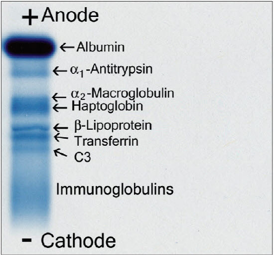
Figure. 2. Positions of major serum proteins in a normal person using electrophoresis in agarose. Individual proteins separate according to their electrical charge between the anode (positive pole) and the cathode (negative pole).
Figure 3. Plasma protein electrophoresis pattern in agarose gel is composed of five fractions, each composed of many individual species. Some of the major proteins are shown here in an artist’s rendition for clarity.
α1Ac, α1-Antichymotrypsin; α1Ag, α1-acid glycoprotein; α1At, α1-antitrypsin; α2-M, α2-macroglobulin; α-Lp, α-lipoprotein; Alb, albumin; AT3, antithrombin III; β-Lp, β-lipoprotein; complement components C1q, C1r, C1s, C3, C4, C5, as designated; C1Inh, C1 esterase inhibitor; Cer, Cerulosplasmin; CRP, C-reactive protein; Gc, Gc-globulin (vitamin D–binding protein); FB, factor B; Fibr, fibrinogen; Hpt, haptoglobin; Hpx, hemopexin; immunoglobulins IgA, IgD, IgE, IgG, IgM, as designated; IαTI, Inter-α-trypsin inhibitor; Pl, plasminogen; Pre A, prealbumin; Tf, transferrin.
Figure 4. Serum protein electrophoresis: clinicopathalogic correlations.
Figure 5. Serum protein patterns in
(1) chronic inflammation with decreased albumin and increased γ-globulins;
(2) acute inflammation with increased α2-fraction (haptoglobin)
and decreased C3 due to activation and consumption of complement;
(3) inanition post–spinal cord injury with hypoproteinemia of several fractions.
Figure 6. Patterns of urine protein electrophoresis in different disorders.
(1) Severe glomerular proteinuria with a major band of albumin plus a secondary one of transferrin (*).
(2) Trace proteinuria with a faint band of albumin and other diffuse proteins.
(3) Immunoglobulin light chains (*).
(4) Tubular proteinuria with multiple bands that do not correspond to major serum proteins.
(5) Hematuria with a major band of hemoglobin
Figure 7. Serum and urine protein electrophoretic patterns in a patient with multiple myeloma. Serum demonstrates a predominance of the larger complete immunoglobulin; the urine has a large amount of the smaller-sized light chains with only a small amount of the whole immunoglobulin.
참고문헌 Henry’s Clinical Diagnosis and Management by Laboratory Methods, 22nd ed.
By Richard A. McPherson, MD and Matthew R. Pincus, MD, PhD (2012)
So you have finished reading the 단백질 전기 영동 결과 해석 topic article, if you find this article useful, please share it. Thank you very much. See more: 전기영동 결과 보는법, 전기영동법, dna 전기영동, Protein electrophoresis, 2차원 전기영동, Immunofixation electrophoresis, 전기영동 종류

